 |
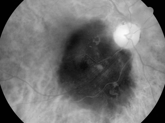
| Figure 7B. Monochromatic photograph of the same fundus taken through a red filter (peak transmission at 615nm) to enhance and document the borders of the lesion |
|
| Figure 7B. Monochromatic photograph of the same fundus taken through a red filter (peak transmission at 615nm) to enhance and document the borders of the lesion |