 |
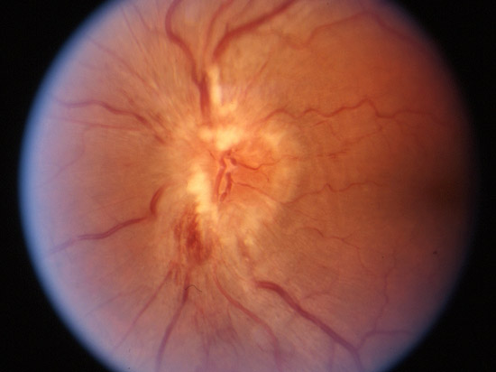
| Figure 11A. Stereo imaging. (A-B) Left and right stereo color fundus photographs of papilledema (swelling of the optic nerve). |
|
| Figure 11A. Stereo imaging. (A-B) Left and right stereo color fundus photographs of papilledema (swelling of the optic nerve). |