 |
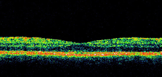
Figure 15A. Optical coherence tomography (OCT). A 3mm line scan provides a cross-sectional view of the normal retinal architecture and foveal depression. |
|
Figure 15A. Optical coherence tomography (OCT). A 3mm line scan provides a cross-sectional view of the normal retinal architecture and foveal depression. |