 |
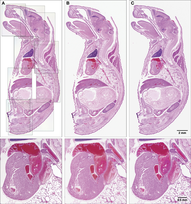
| Figure 2. H&E stained specimen of newborn mouse (specimen courtesy of Dr. Jerrold Ward). A composite image of the specimen captured through traditional photomicrography and a digital microscope camera (images provided by Keith Rogers and Scott Lawrence; some density and color correction applied); B) Specimen scanned directly using an Epson 1640XL flatbed scanner with transparency adapter and Silverfast Ai acquisition software (image provided by Jon Summers; minimal adjustments applied post-acquisition); C)Specimen scanned directly using an Imacon Flextight 949 CCD drum scanner and the process described in the text (minimal adjustments applied post-acquisition). Lower Panels: Enlarged section of the respective panel above showing the heart and a portion of lung. |