 |
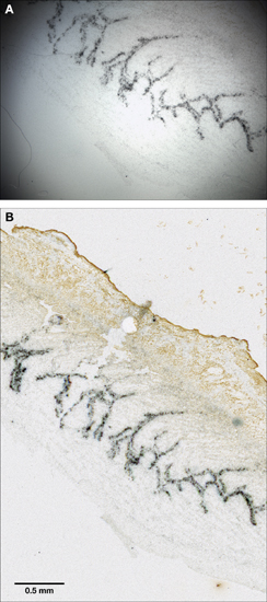
| Figure 5. A) Low-power photomicrograph of luminal epithelium using traditional light microscope and digital camera (specimen and image courtesy of Dr. Clara Rodriguez; some density and color correction applied); B) Same specimen captured using the scanning method described (no image corrections applied; scale matched to panel A). |