|
| Seeing the Body: The Divergence of Ancient Chinese and Western Medical Illustration |
| Camillia Matuk, B.Sc., M.Sc. A medical illustration is the culmination of centuries of medical philosophy, science, and spirituality. Despite similar beginnings, different circumstances surrounding the development of Chinese and Western medical thought eventually led to their divergence. Through an historical comparison, I will attempt to demonstrate how the East and the West came to have such different views, and thus illustrations, of the human body.
|
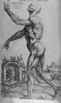
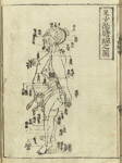
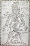
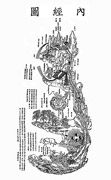
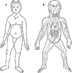
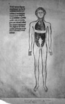
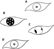
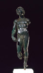
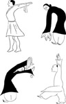
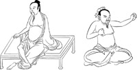
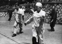
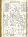
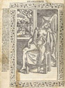
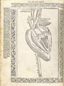
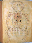
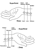
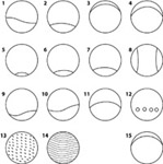
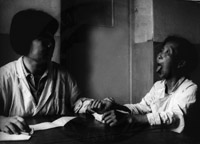
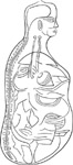
Introduction Seeing, in art as in anatomy, is an acquired skill. Jombert noted that a beginner sees almost no muscles in a nude body (Mayor, 1984). Likewise, Galen said an effort to find muscles must be made in order to observe them (Kuriyama, 1999). But if a Chinese physician saw before him a patient similar to the one a Greek physician saw, what could have led to such different ideas of anatomy? What prevented the Chinese from noticing and even having a word for the elaborations of muscle (Kuriyama, 1999) (Figure 1)? Similarly, what kept the Western physicians from realizing the intricate acupunctural points and tracts that can be delineated across the figure (Figure 2)? This article outlines some major milestones in the development of Eastern and Western medical thought, and provides examples of medical illustrations that reflect their differing philosophies.Similar Beginnings Eastern and Western medicine began with similar fusions of religion, spirituality, and science. Anatomists resorted to analogies of the universe to explain the body when superstitions surrounding death and the fate of the soul prevented closer observation through dissection. To anatomists, nature was divided into elements, each determined by complex associations with gods and all existing by divine will (Mahdihassan, 1973) (Figures 3, 4). Even the diagrammatic anatomy of the Five Pictures Series in Medieval Greece (323 B.C. – 146 B.C.) closely resembles that of The Frog Classic of Huang Di in China's Han period (206 BC - AD220) (Figure 5). Both show contoured human figures, legs and arms splayed and flexed, and laying supine as if on autopsy tables. The accompanying text of the Frog Classic outlines inauspicious dates for cauterization and bleeding; it draws relationships between astrology and blood-letting, subjects that also occupied much of Europe’s pre-Renaissance medical writings (Herrlinger, 1967). Forced to deduce anatomy through speculation and hazy recollections of past experiences, it is no wonder that early anatomical illustrations leaned toward symbolic representations of organs never closely examined (Figures 6, 7). But when the barriers to dissection fell in Medieval Europe (322 B.C.) while remaining erect in China, Eastern and Western medical knowledge diverged. Illustration styles diverged as well: one toward greater realism, and the other preserving the philosophies and thus the symbolism of their earliest studies. What follows is an account of the changing Western medical views of the body, and how illustration developed from the symbolism of The Five Picture Series to the realism of Stephan von Kalkar in Vesalius' De Humani Corpis Fabrica. Greece and the Investigative Spirit Galen, in his book On the Usefulness of the Parts identified three kinds of anatomy students: those who studied physical and mental functions, those who valued the pursuit of knowledge, and those who wished to prove the purposefulness of nature's creations (Kuriyama, 1999). Accordingly, when human dissections were finally sanctioned in Egypt by Alexander the Great (Tarshis, 1969), the impetus was as much a spiritual and aesthetic one as it would eventually become a scientific one. Medicine was not yet advanced enough to rely on the knowledge dissection could provide (Kuriyama, 1999). It was still ruled by Hippocrates' theory of humors, which defined health as a balance between phlegm, blood, yellow bile, and black bile, and did not require support of findings from dissections (Calkins, et. al, 1999). To the Greeks, the body was Nature's masterpiece, each part to be held in awe for the perfection of its form for its purposes. This idea was carried on by Vesalius, who, in his De Humani Corporis Fabrica, praised the Creator for his foresight in constructing the body as it was. Interestingly, much of the interest by European medical illustrators in representing muscles was due to the influence of Western art (Kuriyama, 1999). Since the fifth century B.C. and throughout history, Western art reflected an idea of a perfect body: sinewy and jointed, symbol of vigor and bravery, and a mark of distinctiveness from the masses (Kuriyama, 1999; Physiognomics 810a15-31,). Examples are ubiquitous (Figure 8), even at a time when muscles as an anatomical concept were still considered a tissue yet indistinct from tendons and sinews. Contrast this to the ancient Chinese view of ideal physical health, still evident in modern health practices (Figures 9-11). Here, fluidity of the joints over brute strength was the rule, and the muscle-man of the Fabrica illustrations would have been unimaginable (Kuriyama, 1999). Thus, even before dissection became common practice, Greek anatomists were conditioned by the physical aesthetic of their culture to observing the muscles. However, certain evolutionary milestones had to be reached before achieving the spirit of scientific inquiry that so characterizes Western thought. In examining these events, the importance in the history of Western medicine of preparing the mind to see becomes especially obvious. While dissection had been extensively practiced on animals, the belief in the sanctity of the human body prevented anything but comparative anatomical studies to be drawn, most notably by Aristotle (384-322 B.C.) (Calkins, 1999). Upon Aristotle's death, Egypt became the new hub of intellectual life, and in Alexandria, human dissections finally became sanctioned (Tarshis, 1969). As the first person to base anatomical studies on human dissections, Herophilus of Chalcedon (350-280 B.C.) (Singer, 1925) is credited with a number of contributions; among them: descriptions of the brain, spinal cord, and nerves, and identifying the brain as the body's central organ (contrary to the former Aristotelian belief) (Gordon, 1949). But his extrapolation from animal dissections of the rete mirabile led to belief in a supposed network of vessels permeating the brain. Persistence of this idea through the Middle Ages is proof of the still immature ability to observe with an unbiased eye (Clarke and Dewhust, 1972). With the rise of the Holy Roman Empire over Egypt, a dark age came upon the pursuit of medicine (Lind, 1975). As Roman law forbade human dissection (Singer, 1925), the works of Hellenic Alexandria came to have an overbearing influence on medieval medical thought (Calkins et al., 1999). Great physicians such as Claudius Galen (130-200 A.D.) erroneously assumed similarities between human and animal anatomy, and clung to the theories of 1,500 years before. Even when human dissection was once again permitted in medical schools, it was performed with increasing detachedness of the professor, who sat at a distance and directed a barber through the procedure (Singer, 1925) (Figure 13). Notably, this time also saw the appearance of illustrations accompanying manuscripts of dissections (e.g. John Arderne's De Arte Physicali et de Cirugia, 1412; the anatomical figures in The Fasiculus Medicinae of Johannes de Ketham Alemanus, 1491; and the fugitive-sheets from the 12th century outlining simple anatomy as a study aid for medical students (Calkins et al., 1999)). But while artistic advances were made, the illustrations were often vague. They served only an aesthetic purpose, perpetuating the errors of Galen rather than contributing to scientific progress (Lind, 1975). Dissection was favored more as an artistic pursuit than a scientific one (Ball, 1910) and Hippocrates' and Galen's teachings prevailed even as descriptive anatomical studies through dissection were taking place. The Renaissance brought with it an investigative spirit best captured by Leonardo da Vinci (1452-1519) when he said, "If you find from your own experience that something is a fact and it contradicts what some authority has written down, then you must abandon the authority and base your reasoning on your own findings” (Tarshis, 1969). Later, the anatomist Jacopo Berengario da Carpi (1460-1530) challenged Galen’s teachings, saying "I have never seen this rete (mirabile)" (Clarke and Dewhurst, 1972). Still, anatomical illustration was superfluous to the text, failed to elucidate the structures in question, and often obscured them with unnecessary ornamentation (Figures 14, 15) (Choulant, 1920). It was Andreas Vesalius (1514-1564) who would finally dispel the erroneous notions of Galen and his predecessors (Fulton, 1950). He performed his dissections and employed artists to illustrate his findings with a clarity and didacticism previously unmatched. De Humani Corporis Fabrica, published in 1543, came amid Albrecht Durer's (1471-1528) new considerations of perspective in anatomical illustration (Ficker, 1971), and the developing spirit of scientific inquiry. China and the Alternative Anatomy Although ancient civilizations such as Egypt, China, Babylonia, and India were producing some of history's first medical illustrations before 1500 B.C. (Netter, 1957) (Figure 16), many disregard these as contributions to the development of anatomy (MacKinney, 1965). "It is evident that the Chinese have not pursued in medicine a program calculated to lead them to any great success. They have undoubtedly been held back in this as in other spheres of knowledge by their extreme reverence for ancestral beliefs and customs," (Waye, 1973). Thus, the foundations of anatomical inquiry are traditionally attributed to the Ancient Greeks (Allbutt, 1921). From the Han dynasty (221 B.C. – 220 A.D.) to the 19th century, the West underwent revolutions in printing technology, art, anatomy, and medicine. Disproved theories were continually being replaced with new ones, a revered practice in the West. But, in China, little changed. Rather, history accumulated in layers; new thoughts co-existed with the old ones. Chinese history's quintessential medical text, The Huang Di Neijing (Yellow Emperor's Canon of Internal Medicine), had amassed new theories since before 200 B.C. (Alphen and Aris, 1995) such that the most current medical knowledge always had roots centuries old. Similarly, illustrations remained so unchanged from their earliest symbolic representations that we wonder whether this reflects a lack of the observational skill the Greeks took centuries to perfect. More likely, it shows their concern was not, as it was for the Greeks, over the exact locations and appearances of particular structures; rather, it was on the ideas and deductions to be drawn from them. Ultimately, a chart of acupunctural points and the courses of qi within the body is just as successful in communicating the thought behind it as an illustration from Vesalius' Fabrica is in elucidating tissues such as muscles. Without the means for making concrete observations, the Chinese based their knowledge of anatomy on metaphor. They compared the body to their perceived universe, where health was a balance of Yin (negative, female energy), Yang (positive, male energy), and the Five Phases (earth, water, metal, fire, wood) (Alphen and Aris, 1995). Physicians of China, a country rooted in agriculture, likened the body to a plant. They described a flowering of the face, a body being nurtured by the zang (organs) and illness as a wilting, fading, limpness, shriveling, or desiccation (Kuriyama, 1999). The Chinese drew mystical numerical associations, called the Da shu, or "great numbers.” It was no coincidence to the ancient Chinese, for example, that our four limbs matched the number of seasons and directions, and that in the one record of a human dissection on the body of the rebel Wangsun Qing, the hired butchers of his captor, Wang Mang, reported finding five zang (liver, gall bladder, heart, spleen, kidneys) corresponding to the five planets; 12 vessels circulating blood and air corresponding to the 12 rivers flowing toward the Central Kindgom; and 365 parts of the body, one for each day of the year (Lingshu 13/311). Internal organs were not regarded as distinct entities describable by shape, color or form; or as having distinct functions the way we consider legs suitable for walking and eyes useful for sight. Such things as thought and blood flow were not assigned origins in the brain and in the heart as they had been for the Ancient Greeks (Gordon, 1949). Neither were direct causes and effects acknowledged as when a nerve is cut, the arm falls limp, or when an artery is blocked, the pulse disappears. Rather, Chinese physicians saw unbiased power shifting among the body's parts; they drew indirect causes and effects for affected organs with larger spans of time between events. Thus, a weak spleen could lead to emaciation and a lung injury to a coarsening of the skin (Suwen 8/28 - a manuscript preserving the text of the Nanjing, first compiled during the Han Dynasty 221 B.C. – 220 A.D.). They conceived an imaginary organ system called "the three burning spaces," one of the six fu, distributed over upper, middle, and lower parts of the body, and representing heaven, earth, and man (Veith, 1973). They had mastered the art of pulse diagnosis, well recorded in the Nanjing, (The Classic of Difficult Issues). Pressing the wrist lightly a physician could assess the state of the skin and pores and of the lungs that governed them. Pressing harder, he could determine the state of blood vessels. Still pressing harder, he gleaned information on the tendons and liver, and at the deepest level, he could know the condition of the kidneys and of the bones over which they presided (Figure 17). Illustrations of the pulse, or mo, place it within the theory of the Five Phases; they show links between the hollow pulse of fire, the floating pulse of metal, the slow beat of earth, the deep rhythms of water (Figure 18) (Kuriyama, 1999). What a Greek physician would have manually had to investigate in order to locate the source of illness, the Chinese physician would deduce by a mere look from the five characteristic colours, or wuse, on the body's surface. From these, he could tell whether a patient suffered pain (green or black), cold (white) or fever (red or yellow) (Kuriyama, 1999) (Figure 19). When the Chinese empire became unified and isolated states formed economic ties, the body became a metaphor for the state as well as a microcosm of the universe (Figure 4). It was seen as composed of depots and palaces connected by conduits. Invisible vapors called qi flowed through these conduits and maintained health, while obstruction caused illness (Alphen and Aris, 1995). Acupuncture was a way of influencing the bodily functions by redirecting the flow of qi within the conduits by various techniques of needle insertion. Developed in the 2nd century, it eventually replaced older procedures of bleeding, still a popular practice in the West at the time. Charts of the various directions and locations of qi traditionally show four views: front, back, side, and a view with organs (Alphen and Aris, 1995). These charts, created during the seven centuries between the Song period and the 19th century, demonstrate the ancient Chinese disregard for specific organ morphology (Figure 20). Conclusion Dissections in Ancient Greece, although they contributed greatly to knowledge of anatomy in the West, were not enough to dispel the misconceptions in medicine's past. Even as Greek physicians, and on one occasion, Chinese physicians, effectively examined the same internal structures, both came to define anatomy in different ways. The illustrations shown in this article represent a culmination of centuries of philosophy, reason, and deduction. Just like Jombert’s artists, Western anatomists had to first conceive of the idea of muscles before they would ever come to notice them. The elaborate musculature in Vesalius' Fabrica demonstrates their prepossession for physical minutiae. Meanwhile, in their more holistic philosophies on health and disease, the Chinese found no reason to even imagine a muscle present in the body, and thus, they saw none. Rather, the iconized figures in their illustrations suggest alternative ways of understanding anatomy; intangible ways that connected their selves to their universe. References Allbutt, T. C. 1921. Greek medicine in Rome. London: Macmillan, 633. Alphen, J. V., Aris, A. eds. 1995. Oriental Medicine: An illustrated guide to the Asian arts of healing. London, England: Serindia Publications, 159. Ball, J. M. 1910. Andreas Vesalius, the reformer of anatomy. St Louis: Medical Science Press. p.149. Calkins, C. M., J. P. F ranciosi, and G. L. Kolesari. 1999. Human anatomical science and illustration: The origin of two inseparable disciplines. Clinical Anatomy 12: 120-129. Choulant, L. 1920. History and bibliography of anatomic illustration in its relation to anatomic science and the graphic arts. Chicago: University of Chicago Press, 435. Clarke, E., Dewhurst, K. 1972. An illustrated history of brain function. Berkeley: University of California Press, 154. Ficker, F. 1971. Albrecht Dürer and the medicine of his time. Munch Med Wochenschr 113: 814-818. Fulton, J. F. 1950. Vesalius four centuries later: Medicine in the eighteenth century. Lawrence: University of Kansas Press, 52. Gordon, B.L. 1949. Medicine throughout antiquity. Philadelphia: Davis, 818. Herrlinger, R. 1967. Geschichte der medizinischen Abbildung (History of Medical Illustrations). Munich: Heinz Moos Verlag, 30. Kuriyama, S. 1999. The expressiveness of the body and the divergence of Greek and Chinese medicine. New York, N. Y.: Zone Books. Lind, L. R. 1975. Studies in pre-Vesalian anatomy. Philadelphia: American Philosophical Society, 344. MacKinney, L. 1965. Medical illustrations in medieval manuscripts. Los Angeles: University of California Press, 262. Mahdihassan, S. 1973. A rational interpretation of the four cosmic elements as operating in alchemy. In Theories and philosophies of medicine. 2nd ed. Compiled by Department of Philosophy of Medicine and Science. Tughlaqabad, New Delhi 62: Institute of History of Medicine and Medical Research. Mayor, A. Hyatt. 1984. Artists and Anatomists. New York: Artists Limited Edition, 10. Netter, F. 1957. Medical illustration – Its history, significance and practice. Bull New York Academy of Medicine 33: 357-368. Singer, C. 1925. The evolution of anatomy: A short history of anatomical and physiological discovery to Harvey. New York: Alfred A. Knopf, 209 Tarshis J. 1969. Father of modern anatomy: Andreas Vesalius. New York: Dial,103. Veith, I. 1973. Universalistic concepts in Chinese anatomy and medicine. In Theories and philosophies of medicine. 2nd ed. Compiled by Department of Philosophy of Medicine and Science. Tughlaqabad, New Delhi 62: Institute of History of Medicine and Medical Research. Waye, J.D. 1973. A short account of Chinese medicine. In Theories and philosophies of medicine. 2nd ed. Compiled by Department of Philosophy of Medicine and Science. Tughlaqabad, New Delhi 62: Institute of History of Medicine and Medical Research. Werner, E.T.C. 1958. Myths and Legends of China. London: George G. Harrap & Co. Ltd., 184-188. Werner, E.T.C. 1969. A Dictionary of Chinese Mythology. New York: The Julian Press, Inc., 43, 320. Acknowledgements For the two years prior to receiving a B.Sc. in Biological Sciences from the University of Windsor (2002), Camillia Matuk lived and studied in Hong Kong. In 2004, she earned a M.Sc. in Biomedical Communications from the University of Toronto and a post-graduate diploma in 3D Computer Animation from the Sheridan College Institute of Technology and Advanced Learning. Until 2006, she was a medical illustrator at the Toronto-based medical animation studio, InViVo Communications, Inc. Currently she is working toward a Ph.D. in The Learning Sciences at Northwestern University in Evanston, Illinois. |
Copyright
2006, The Journal of Biocommunication, All Rights Reserved
Table
of Contents for VOLUME 32, NUMBER 1