 |
||||
Book Review |
Jim Perkins
|
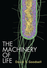
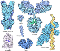
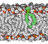
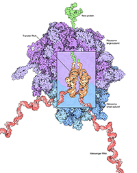
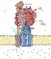
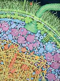
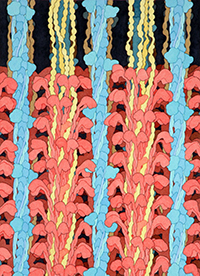
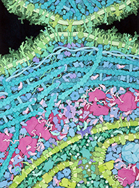
The Machinery of Life, 2nd Edition by David S. Goodsell, PhD. Published by Copernicus Books, an imprint of Springer Science and Business Media, 2009. 167 pages. More than fifteen years after its initial publication, the long-awaited second edition of David Goodsell’s The Machinery of Life was released in 2009. This is a wonderful introduction to the world of molecules and cells for the nonscientist, but also serves as a handy refresher and an enjoyable read for those already well versed in molecular and cell biology. David Goodsell is Associate Professor of Molecular Biology at The Scripps Research Institute, where he works in their Molecular Graphics Laboratory. He is also an accomplished scientific illustrator, using 3D molecular graphics and his own unique style of watercolor painting to bring molecular and cellular subjects to life. He has presented at the Association of Medical Illustrators annual meeting, exhibited work in the Guild of Natural Science Illustrators members exhibition, and has contributed to this journal (Goodsell 2000, JBC 27[4]: 12-18). He writes and illustrates the Molecule of the Month feature at the Protein Databank and has written three books: The Machinery of Life (1st ed. 1993), Our Molecular Nature: The Body's Motors, Machines, and Messages (1996), and Bionanotechnology: Lessons From Nature (2004), all profusely illustrated in his unique style. In 1999, he received the Frank H. Netter Award for Special Contributions to Health Science Education for the first edition of The Machinery of Life. For those of you already familiar with this book, the most obvious change in the second edition is that all of the illustrations are now in full color. It’s now possible to distinguish atoms based on the familiar CPK color scheme (red oxygen, blue nitrogen, etc.). The use of color also makes it far easier to distinguish neighboring structures in Goodsell’s cellular paintings, which emphasize the dense crowding of molecules inside the cell. The illustrations in the second edition are not simply colorized versions of old figures. Nearly every illustration appears to have been created from scratch, including computer generated 3D models and the watercolor paintings of cellular interiors. The text has also been largely re-written and incorporates the latest research in biochemistry and cell biology. There is an entirely new chapter on “Life and Death” that describes (and illustrates) recent discoveries regarding programmed cell death (apoptosis), DNA repair, the important role of the telomere, and the processes of cancer, aging, and death. Chapter 1 begins with an excellent discussion of scale, comparing the sizes of molecules, cells, and whole organisms. This is useful for the illustrator who must sometimes depict structures at different scales in the same figure. Of course, the differences in scale between a single molecule and an entire cell are so vast that the illustrator must often use artistic license to depict them in the same image. Nevertheless, it is useful to understand these differences in scale so the illustrator knows just how much “license” he/she is taking in order to make the figure work. Goodsell has wisely adopted a consistent scale for most of his illustrations. All of the hand-painted cellular landscapes are shown at 1,000,000x. The illustrations of individual molecules are somewhat more variable – most are at 5,000,000x with some close-ups at 20,000,000 to 40,000,000x. The magnification is included in each figure legend, making it easy to compare one illustration with another. Chapter 2 introduces the concept of molecules as machines, including a brief overview of the four major classes of biological molecules – nucleic acids, proteins, lipids, and polysaccharides. Chapter 3 – The Processes of Living – describes how organisms construct new components from basic building blocks, harness energy from their surroundings, regulate the influx and efflux of materials, and perceive and respond to their environment. Chapters 4–6 explore these processes in more detail, using two organisms as examples: the bacteria Escherichia coli (Ch. 4) and the human (Chs. 5 and 6). The latter chapters also discuss the advantages of compartmentalized organelles found in the eukaryotic cell and the specialization of cells in multicellular organisms. Chapter 6 includes detailed descriptions of muscle, blood, and nerve tissue as examples of cellular specialization. The remaining three chapters describe important molecular interactions between the body and its environment. Chapter 7 – Life and Death – describes the recycling of damaged proteins in the cell, DNA repair, free radical damage, apoptosis, and the inevitable processes of aging and death. Chapter 8 describes four different viruses (poliovirus, rhinovirus, influenza, and HIV) and how they infect human cells, while the final chapter – You and Your Molecules – discusses the role of vitamins in the body and the effects of poisons, bacterial toxins, and drugs. For those interested in molecular modeling, Goodsell includes an appendix with a list of all Protein Databank files used in creating his molecular models. The single greatest strength of The Machinery of Life is the high quality of its illustrations. Goodsell developed his own molecular graphics software while doing post-doctoral work with Art Olson in Scripps’ Molecular Graphics Lab. His molecular images use a variation on the space-filling or CPK style where each atom is rendered as a sphere, proportional in size to the atom’s van der Waals radius. His images lack the bright highlights and shiny textures so common in molecular illustrations. Instead, his molecules are rendered with flat colors and black outlines, yet the use of shadows still gives them a strong sense of depth and form. In his own words, “I like the way that this style simplifies the molecule, giving a feeling for the overall shape and form of the molecule, but at the same time you can still see all the individual atoms. (PDB Newsletter 2003, p. 5).” The space-filling style also facilitates the representation of molecules at different levels of magnification. Again in the Goodsell’s own words: “The use of a space-filling approach allows the combination of close-up pictures, which show the atomic details of each molecule, with larger fields of molecules, where atomic detail would add too much complexity. By progressively smoothing the representation as larger and larger fields are shown, the image maintains an appropriate level of comprehensibility at each scale level. The similarity in shape between the different levels allows the viewer to move from one image to the next and identify individual molecules (Goodsell 2005, p. 353).” Goodsell’s large-scale paintings of cellular contents are particularly impressive. He has single-handedly changed the way cells are perceived. They are not giant water balloons, sparsely populated with tiny molecules. They are jam-packed with large molecules, such as proteins and nucleic acids, which occupy 25-35% of the available space (to say nothing of the myriad small molecules dissolved in the cytoplasm). Molecules in the cell interact with one another, not by whizzing through an aqueous void, but by bumping and jostling like passengers on a crowded subway train. Goodsell’s paintings show every macromolecule “at the proper size and shape, at the proper concentration, and in the proper location” within the cell (Goodsell 2000, p. 18). Goodsell’s writing is just as clear and enjoyable as his artwork, as evidenced by this passage about red blood cells: “Red blood cells are unselfishly dedicated to their work of carrying oxygen from the lungs to the tissues. In fact, they can do little else. Red blood cells are created from stem cells in the bone marrow. As they develop, they gradually shift their resources almost entirely to the building of hemoglobin, and allow all of their other functions to atrophy. The cell membrane loses much of its machinery for communication and selective transport, and is braced only by a rudimentary scaffolding that helps the cell hold its distinctive disk-like shape. Finally, the cell makes the ultimate sacrifice. It concentrates all of its normal molecular machinery – mitochondria, nucleus, ribosomes – into one corner and ejects it all from its body. The mature red blood cell, now a directionless automaton, is then placed in the blood-stream... (p. 94).” No book review is complete without at least a little bit of constructive criticism, lest the reader conclude that I’m on the Scripps payroll. The book includes a handful of typos, something that’s probably inevitable in the first printing of any book. For example, on page 103, Goodsell writes: “other synapses, such as those in the central nervous signal, send weaker messages that involve only a single vesicle.” Presumably he meant “central nervous system”. There’s also inconsistency in the capitalization of “cytochrome P450”. It appears as P450 on one page and p450 on another. These are all minor issues that may already be fixed in preparation for a second printing. A more significant issue is the format of Chapter 5 – A Human Cell. After a brief 1-1/2 page introduction, the remainder of the chapter consists entirely of illustrations and their accompanying figure legends. There is no text. This might sound like an odd criticism coming from an illustrator – you might think I’d welcome a chapter full of artwork – but it’s inconsistent with every other chapter in the book. It just seemed a bit odd, as if the publisher asked him to trim five or six pages just prior to publication. I would have welcomed some additional explanatory text in this chapter. Finally, I had difficulty locating and reading the labels in many of the cellular paintings. Rather than labeling these figures with whole words, Goodsell placed a single black letter on each molecule that referred to a key in the figure legend. This was a good choice since whole words would have been impossible to read against the densely cluttered cellular landscapes. However, the individual letters get lost amidst the complexity of the images. I spent considerable time searching for some of them (imagine “Where’s Waldo” in the molecular realm). This problem is exacerbated by the fact that many of the paintings are fairly dark and every molecule is outlined with a black stroke. Bold white letters may have been a better choice against the darkly colored backgrounds. Despite these minor concerns, I highly recommend The Machinery of Life for both the educated lay reader and for the scientist. As Goodsell states in the preface, “I have written the text with the nonscientist reader in mind, and I have drawn the illustrations at a level of scientific rigor meant to satisfy readers who are scientists.” For the scientific illustrator, the artwork alone is well worth the price of the book.
References Goodsell, D.S. 1993. The Machinery of Life, 1st ed. NY: Springer-Verlag. Goodsell, D.S. 1996. Our Molecular Nature: The Body's Motors, Machines, and Messages. NY: Springer-Verlag. Goodsell, D.S. 2000. Illustrating the 'Machinery of Life'. Journal of Biocommunication 27(4): 12-18. Goodsell, D.S. 2004. Bionanotechnology: Lessons From Nature. NY: Wiley-Liss. Goodsell, D.S. 2005. Visual methods from atoms to cells. Structure 13(3): 347–354. RCSB Protein Databank. 2003. PDB Focus: David Goodsell and the Molecule of the Month. PDB Newsletter No. 17, Spring 2003.
Jim Perkins, MS, MFA, CMI, FAMI, is Associate Professor of Medical Illustration at Rochester Institute of Technology and Adjunct Instructor in Anatomy for the Ithaca College School of Health Sciences and Human Performance. He is a Board Certified Medical Illustrator and Fellow of the Association of Medical Illustrators. Jim has a special interest in molecular illustration and has given talks and workshops on the subject for the Association of Medical Illustrators and the Guild of Natural Science Illustrators. He received the 2006 AMI Literary Award for his two articles on the history of molecular visualization (JBC 31-1 and JBC 31-2). japfaa@rit.edu
Editor's Note See more illustrations by Dr. Goodsell in his feature article, Getting the Most Out of the Protein Data Bank.
|
||||||||
|
Copyright
2010, The Journal of Biocommunication, All Rights Reserved |